Flavonoid-Rich Cocoa Extract Alleviates Oxidative Stress, Inflammation and Liver Dysfunction Induced by High-Fat Diets in Wistar Rats
-
Igbayilola Yusuff Dimeji


Department of Human Physiology, College of Medicine and Health Sciences, Baze University, Abuja, Nigeria
Grema Mariam GujjaDepartment of Human Physiology, College of Medicine and Health Sciences, Baze University, Abuja, Nigeria
Saka Waidi AdeoyeDepartment of Physiology, Faculty of Basic Medical Sciences, Ladoke Akintola University of Technology, Ogbomoso, Nigeria
Hamidu, Lawan JabbaDepartment of Human Physiology, College of Medicine and Health Sciences, Baze University, Abuja, Nigeria
Jibrin, SherifdeenDepartment of Biochemistry, Faculty of Computing and Applied Sciences, Baze University, Abuja, Nigeria
Zakari Muhammed BabaDepartment of Human Physiology, College of Medicine and Health Sciences, Baze University, Abuja, Nigeria
| Received 09 Jul, 2024 |
Accepted 11 Oct, 2024 |
Published 12 Oct, 2024 |
Background and Objective: The escalating prevalence of metabolic disorders associated with high-fat diets emphasizes the necessity for effective nutritional interventions. Cocoa flavonoid-rich aqueous extract (CHFAE), known for its polyphenolic richness, emerges as a promising therapeutic option. This study investigates CHFAE's impact on antioxidant, inflammatory, and liver enzyme bio-markers in Wistar rats subjected to a 12-week high-fat diet. Materials and Methods: Rats were grouped into a normal diet (Group 1), normal diet with 600 mg/kg cocoa flavonoid extract (Group 2), a high-fat diet with 600 mg/kg cocoa flavonoid extract (Group 3), and an untreated high-fat diet (Group 4). CHFAE was administered orally for 14 days. After the period of the experiment, biomarkers of oxidative stress such as SOD, CAT, GSH, and lipid peroxidation’s index MDA were assayed. Also, liver function parameters such as AST, ALT, and ALP were quantified, and inflammatory cytokines such as IL-6, and TNF-α were assayed. The data were presented as mean ± standard error. One-way ANOVA was employed to assess statistical differences between groups, followed by Tukey's post hoc test for individual comparisons. A 95% confidence interval was applied, and results with P < 0.05 were deemed statistically significant. Results: Results showed a significant increase in GSH, SOD, and CAT levels and a concomitant decrease in lipid peroxidation’s MDA index compared to controls and untreated high-fat diet rats. Inflammatory cytokines (IL-6, TNF-α) and liver function biomarkers (AST, ALT, ALP, albumin, total protein) showed significant reduction in CHAFE-treated rats compared with control. Conclusion: This study suggests that CHFAE may effectively combat oxidative stress, inflammation, and liver dysfunction caused by high-fat diets, encouraging further exploration for potential health-promoting functional foods or supplements.
INTRODUCTION
In recent years, the overall prevalence of obesity and related metabolic disorders has reached alarming levels, which is a major public health challenge1. Lifestyle factors, including dietary habits, play a crucial role in the development and progression of these diseases. High-fat diets, characterized by excessive consumption of saturated fat and refined sugars, promote oxidative stress, inflammation and liver dysfunction2. As a result, researchers have increased their efforts to identify natural compounds that have potential therapeutic effects against these adverse effects. One such compound under study is a flavonoid-rich aqueous extract of cocoa, known for its antioxidant and anti-inflammatory properties.
Cocoa, derived from the Theobroma cacao plant, has received considerable attention for its potential health benefits. Cocoa is rich in bioactive compounds, especially flavonoids, which are known for their antioxidant properties3. Flavonoids act through several mechanisms, including modulation of glucose metabolism and reduction of oxidative stress. In addition, cocoa consumption has been associated with several health benefits, including improved insulin sensitivity, lipid profile and regulation of blood pressure4. Cocoa flavonoids, particularly epicatechin and catechin, have been widely studied for their potential anti-diabetic and antioxidant properties. These bioactive compounds have demonstrated their ability to improve insulin sensitivity, regulate glucose metabolism and alleviate oxidative stress in both animal models and human studies5,6. Additionally, cocoa consumption has been associated with a decreased risk of cardiovascular disease and improved endothelial function7,8.
Oxidative stress, which results from an imbalance between the production of reactive oxygen species (ROS) and the body’s antioxidant defense mechanisms, is a hallmark of metabolic disorders. Cocoa flavonoids are known for their strong antioxidant activity, acting as scavengers of free radicals and modulators of the redox state of cells9. These properties make cocoa an interesting candidate for ameliorating oxidative stress associated with a high-fat diet. A study by Lee et al.5 and Caminiti et al.6 showed that cocoa flavonoids effectively reduced oxidative stress markers such as malondialdehyde (MDA) and superoxide dismutase (SOD) activity in Wistar rats fed a high-fat diet. The study highlighted the potential of cocoa flavonoids to restore redox balance and protect against cellular damage caused by oxidative stress. Chronic low-grade inflammation is a key factor in the progression of metabolic disorders and a high-fat diet contributes significantly to the inflammatory environment. Cocoa flavonoids have been studied for their anti-inflammatory potential and research suggests modulation of inflammatory mediators and pathways9. Pro-inflammatory cytokines, including interleukin-6 (IL-6) and tumor necrosis factor-alpha (TNF-α), were reduced in adipose tissue of Wistar rats supplemented with cocoa aqueous extract rich in flavonoids. Animal studies provide an excellent opportunity to assess the physiological effects of cocoa and the contribution of cocoa consumption to various models of inflammation. Recent chronic studies by Goya et al.3 and Asiedu-Gyekye et al.10 in which rats received 5 and 12% cocoa powder, respectively found no signs of inflammatory processes or carcinogenicity. Understanding the potential benefits of cocoa, a flavonoid-rich aqueous extract, in ameliorating oxidative stress and inflammation induced by a high-fat diet is crucial for developing treatment strategies for obesity-related complications. This study aims to fill existing knowledge gaps by investigating the mechanisms underlying the antioxidant and anti-inflammatory properties of cocoa flavonoids in a relevant animal model. The findings may inform the development of new nutritional interventions or supplements to combat the adverse effects of a high-fat diet on metabolic health.
MATERIALS AND METHODS
Study duration: This study was conducted between July to October, 2023.
Cocoa seed: In Isemi-Ile, Oyo State, Nigeria, a local farmer named Alhaji Prince Abdur-Rasaq Igbayilola supplied us with 2 kg of dried cocoa seed. Before being extracted, the cocoa seed was ground into a powder using an industrial grinding machine (Foss Analytical, Denmark)11. At the Department of Life Sciences at Baze University Abuja, dried cocoa seed was recognized. A voucher number, BUHOOOO105, was provided and the specimen was placed in the university’s herbarium.
Ethical approval: The ethical approval was sought and given after the proposal presentation to conduct this study from the Department of Human Physiology, Baze University, Abuja, Nigeria.
Extraction
Preparation of standardized cocoa extract: According to Igbayilola and Gujja11, cocoa extract was prepared by extracting defatted cocoa powder with 80% distilled water for 2 hrs. Water was removed using a rotary evaporator (Büchi Labortechnik AG, Switzerland) for 40 min at 55°C. The resulting extract was stored at -80°C and lyophilized. For standardization, 5 mL of the extract was fractionated on a pre-packed column. The elution medium’s water-to-acetone ratio was progressively increased (85:15, 70:30 and 40:60) with a flow rate of 2.0 mL/min. The resulting fractions (F1, F2 and F3) were used to identify bioactive compounds. Fine-tuning separation parameters with acetone improved the chromatography process.
Flavonoid content in cocoa extract: The aluminium chloride colorimetric approach was utilized to ascertain the flavonoid content present in the specimen12. Using this method, flavonoids react to generate a less stable salt complex at the ortho-hydroxyl groups on rings A and B and a more stable acid complex at the C-4 position and the hydroxyl groups at C-3 or C-5. With a few minor adjustments, the flavonoid content of cocoa peel was measured using the techniques of Bag et al. 12. The 20, 40, 60, 80 and 100 μg/mL of quercetin standards were used in an external calibration.
Animals: In this investigation, twenty-four male Wistar rats weighing between 80 and 100 g were employed. The Human Physiological Animal House at Baze University in Abuja served as the venue for them. Wistar rats were housed in clear rat enclosures with a 12 hrs artificial light/dark cycle. The experimental technique was started after two weeks of acclimatization.
Ethical consideration: The Ethics and Review Committee of the Department of Human Physiology at Baze University in Abuja was consulted to obtain ethical permission to perform this study after successful proposal presentation.
Estimation of alkaloids: A separate 250 mL beaker was filled with around 5 g of powdered cocoa and 200 mL of 20% acetic acid. After covering the beaker, it was let to stand for 4 hrs. After filtering the mixture containing the solution, the volume was reduced to one-quarter using a water bath. This sample was treated drop by drop with concentrated ammonium hydroxide until the precipitate was completely produced. The precipitate was filtered out and weighed after allowing the mixture to settle completely13. As a result, the percentage of total alkaloid content was as follows:
Estimation of total phenolic content: The total phenolic content of cocoa was determined using the Folin-Ciocalteau reagent, following the method by Makkar et al.14. Approximately 20 μg of cocoa extract was dissolved in 1 mL of distilled water, followed by the addition of 2.5 mL of 20% sodium carbonate (Na2CO3) and 500 μL of diluted Folin-Ciocalteau reagent (1:1 with water). The mixture was thoroughly mixed and left in the dark for 40 min to develop color. Absorbance was measured at 725 nm. A calibration curve with gallic acid showed linearity between 10 and 50 μg/mL. Total phenolics were expressed as mg gallic acid equivalent (mg GAE/g extract).
Estimation of total flavonoid content:The total flavonoid content was determined following the method outlined by Bag et al.12. To 1 mL of diluted cocoa extract, 150 μL of 5% sodium nitrite solution was added, followed by a 5 min incubation. Next, 150 μL of a 10% aluminium chloride solution was added and allowed to stand for 6 min. Subsequently, 2 mL of a 4% sodium hydroxide solution and 5 mL of distilled water were added. The mixture was shaken and left at room temperature for 15 min. Absorbance was then measured at 510 nm. A pink color indicated the presence of flavonoids, quantified as rutin equivalent mg RE/g extract on a dry weight basis using a standard curve.
Estimation of tannin content: Cocoa's tannin content was assessed using the method by Makkar et al.14. First, 500 μL of extract was mixed with 500 μL of distilled water and 100 mg of polyvinyl polypyrrolidone in test tubes. The mixture was incubated at 4°C for 4 hrs. After centrifuging for 5 min at 5000 r/min, 20 μL of the supernatant, containing only simple phenolics, was collected. Tannins precipitate with polyvinyl polypyrrolidone. The free phenolics in the supernatant were measured at 725 nm and calculated on a dry matter basis. The tannin content of the extract was then determined using a specific formula:
Estimation of total saponin content: Using a modified version of Makkar et al.14, total saponins were estimated based on the vanillin-sulphuric acid colorimetric reaction. To 250 μL of distilled water, we added 50 μL of cocoa extract, followed by 250 μL of vanillin reagent (800 mg vanillin in 10 mL 99.5% ethanol). Next, 2.5 mL of 72% sulfuric acid was added and thoroughly mixed. This mixture was heated at 60°C in a water bath for 10 min, then cooled in ice water. Absorbance was measured at 544 nm. The saponin content, expressed as diosgenin equivalents (mg DE/g extract), was determined using a standard curve.
Estimation of reducing sugar: Fehling’s test also referred to as Benedict's test, was utilized to determine the reduced sugar content in cocoa. When reducing sugars such as glucose, fructose and maltose react with copper ions in the alkaline solution, the color changes from blue to brick-red or yellow, depending on the amount of reducing sugars present15.
Estimation of steroids: High-performance Liquid Chromatography (HPLC) was employed to identify and quantify steroids in cocoa. This technique separates and identifies components based on their chemical properties and retention times.
Estimation of cardiac glycosides: One method for detecting cardiac glycosides in cocoa is the Kedde reagent test. In this test, the sample suspected of containing cardiac glycosides is combined with the Kedde reagent, a mixture of ferric chloride and glacial acetic acid. A distinctive color change, typically from yellow to green or violet, indicates the presence of cardiac glycosides16.
Induction of obesity in Wistar rats: The high-fat diet (HFD) was prepared following the methods described by Amin and Nagy17. Obesity was induced in rats using an HFD consisting of 49% fat (from ghee and corn oil), 32% carbohydrates and 19% protein. In contrast, normal rats received standard chow with 14% fat, 61% carbohydrates and 25% protein. Rats in groups 3 and 4 were fed the HFD for 12 weeks. Body weight was monitored weekly to confirm obesity development and body mass index (BMI) was calculated by dividing body mass (g) by the square of the length from nose to anus (cm).
Treatment regimen: The standardized flavonoid-rich (600 mg/kg body weight) cocoa extract was given to high-fat fed rats orally for 2 weeks (week 13 to15) and grouped thus according to Igbayilola and Gujja11:
| Group 1: Negative control (Received normal rat diet only throughout the period of experiment) | ||
| Group 2: Positive control treated (Received normal rat diet and extract at 600 mg/kg body weight) | ||
| Group 3: Untreated fat (Received high fat diet and extract at 600 mg/kg body weight) | ||
| Group 4: Fat treated (Received high fat diet only throughout experiment) |
Flavonoid-rich cocoa extract was dissolved in 3 mL of 0.03% (w/v) low condensed fat milk (LCFM) and was given daily by oral administration, using an oral cannula. Rats in the control group received 0.03% (w/v) normal saline only throughout the experimental period.
Collection of blood sample: Rats were humanely euthanized under sedation with ketamine. Blood was collected via cardiac puncture into plain tubes, left to clot at 40°C for 1 hr and then centrifuged at 3000×g for 15 min to separate serum samples for biochemical analysis. The serum samples were stored at -20°C until they were assayed11.
Collection of tissue sample: The animals were dissected and their livers and tissues were carefully removed, washed in ice-cold trichloroacetic acid, rinsed with 1.15% KCl solution and then blotted dry.
Homogenization: All collected tissues were homogenized in ice-cold 0.1M phosphate-buffered saline (pH 7.4) and a 10% homogenate was prepared. The homogenate was then centrifuged for 15 min at 3000×g and the resulting supernatant was carefully poured off and stored at -40°C until further analysis.
Liver functions: Albumin, total protein, alkaline phosphatase (ALP), alkaline aminotransferase (ALT) and aspartate aminotransferase (AST) were determined using liver homogenate samples by an automated analyzer (Mindray BS-120, Chema Diagnostica, Italy).
Superoxide dismutase (SOD): The SOD activity was assayed according to the method described by Vilela et al.18.
Reduced glutathione (GSH): The GSH activity was assayed as described by Vilela et al.18. To estimate GSH levels, 50 mL of plasma was combined with 1.78 mL of 0.1 M pH 8 phosphate buffer and 0.2 mL DNTB, then allowed to sit for one hour. The amounts of plasma GSH were then measured using spectrometry on the mixture at a wavelength of 412 nm. For reference, the usual range is 3.8-8.55.
Analysis of catalase (CAT) activity: The usual method was used to estimate the CAT activity. Each treated group’s 1 mL of brain homogenate was added to 5 mL of phosphate buffer (pH 7.0) and 4 mL of H2O2 solution (800 μmol) was then mixed in. This was combined with a gentle spin of the mixture at room temperature. Then, using a spectrophotometer (American-based Agilent Technologies is the manufacturer of the spectrophotometer), a part of the reaction-roughly 1 mL-was collected and given to 2 mL of dichromate/acetic reagent. The changes in absorbance were then recorded at 570 nm and the process was then transformed into catalase activity according to Umukoro et al.19.
Malondialdehyde (MDA): The MDA level was assayed according to the protocol described by Umukoro et al.19. Absorption at 532 nm was compared to an MDA standard curve derived from the hydrolysis of 1,1,3,3-tetramethoxypropane under acid catalysis to determine the reactive components of thiobarbituric acid. Then 0.25N hydrochloric acid, 15% trichloroacetic acid and 0.375 percent thiobarbituric acid were combined to create the mixture. After boiling for 10 min, each sample was mixed with 500 μL of solution and 250 μL of serum, then centrifuged at 3000×g rpm for 10 min. The 200 μL of each supernatant were added to microplates and optical density was measured at 535 nm to express MDA as μmol/L.
Assay of tumour necrosis factor alpha (TNF-α): The Enzyme-linked Immunosorbent Assay (ELISA) test was employed in the assay of serum TNF-α with the aid of a rat TNF-α ELISA kit (Elabscience, Wuhan, China). Cat No: E-El-R2856.
Principle: A suitable quantity of coated strips were inserted into the holder. Pipetting 50 mL of IL-6 standards, control and specimens into the designated wells was done. For 10 to 30 sec, 100 mL of the conjugate reagent-which was now ready for use-was added to each well. The plate was covered and left to stand at room temperature (18-26°C) for 60 min. They took liquid out of each well. Each well received 100 mL of TMB substrate, which was then incubated for 15 min at room temperature. To every well, 50 mL of stop solution was added. A gentle shake of the plate was used to mix the fluid. After adding the stopping solution and waiting for 15 min, an ELISA reader set (Bio-Rad Laboratories, USA) was used to measure the absorbance.
Assay of interleukin-6 (IL-6): The Enzyme-linked Immunosorbent Assay (ELISA) test was employed in the assay of serum IL-6 with the aid of a rat IL-6 ELISA kit (Elabscience, Wuhan, China). Cat No: E-El-R0015.
Principle: A suitable quantity of coated strips were inserted into the holder. Pipetting 50 mL of IL-6 standards, control and specimens into the designated wells was done. For 10 to 30 sec, 100 mL of the conjugate reagent-which was now ready for use-was added to each well. The plate was covered and left to stand at room temperature (18-26°C) for 60 min. Then took liquid out of each well. Each well received 100 mL of TMB substrate, which was then incubated for 15 min at room temperature. To every well, 50 mL of stop solution was added. A gentle shake of the plate was used to mix the fluid. Following the addition of the stopping solution, an ELISA reader (Bio-Rad Laboratories, USA) was used to measure the absorbance after 15 min of waiting.
Statistical analysis: The data were presented as Mean±Standard Error. One-way ANOVA was employed to assess statistical differences between groups, followed by Tukey’s post hoc test for individual comparisons. A 95% confidence interval was applied and results with p<0.05 were deemed statistically significant (GraphPad Prism 5, GraphPad Software, Inc., La Jolla, CA, USA).
RESULTS
Phytochemical analysis: Table 1 shows the phytochemical screening of powdered cocoa beans and the phytochemicals present are as presented.
Influence of flavonoid-rich cocoa aqueous extract on GSH, SOD, CAT activities and MDA level
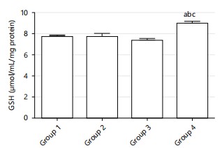
Glutathione (GSH) activity: The oxidative stress study results revealed a significant increase in GSH activity (p<0.05) in both group 2 and 4 when compared to the control (CONT) and group 3. Conversely, the GSH activity in group 3 did not show a significant difference (p<0.05) compared to CONT. Additionally, GSH activity was significantly reduced (p<0.05) in group 3 compared to group 2, while no significant difference (p<0.05) was observed between group 2 and 4. These findings suggest that the cocoa flavonoid aqueous extract may enhance GSH activity, potentially contributing to improved antioxidant defense mechanisms as shown in Fig. 1.
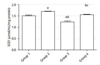
Superoxide dismutase (SOD) activity: The results indicated a significant increase (p<0.05) in SOD activity in groups 2 and 4 compared to the control group (CONT). In contrast, SOD activity in group 3 was not significantly different (p<0.05) from the control group. Furthermore, SOD activity in group 3 was significantly lower (p<0.05) than in group 2, with no significant difference (p<0.05) between group 2 and 4. This suggests that the cocoa flavonoid aqueous extract may enhance SOD activity, thereby potentially reducing oxidative stress as shown in Fig. 2.
| Table 1: | Qualitative phytochemical analysis of aqueous extract of powdered cocoa beans | |||
| Component | Inference |
| Saponin | + |
| Tannin | + |
| Reducing sugars | + |
| Flavonoids | + |
| Alkaloids | + |
| Steroids | + |
| Cardiac glycosides | + |
| Phenols | + |
| +: Present | |
|
|
|
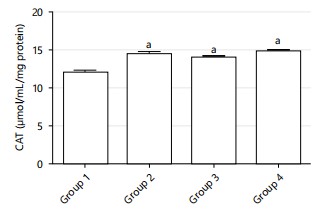
Catalase (CAT) activity: The study results demonstrated a significant increase (p<0.05) in CAT activity in group 2 and 4 compared to the control group (CONT). However, CAT activity in group 3 did not show a significant difference (p<0.05) compared to the control. Additionally, CAT activity in group 3 was significantly lower (p<0.05) than in group 2, while no significant difference (p<0.05) was observed between group 2 and 4. These findings indicate that the cocoa flavonoid aqueous extract might enhance CAT activity, which could be beneficial in neutralizing harmful hydrogen peroxide in cells as shown in Fig. 3.
|
|
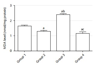
Malondialdehyde (MDA) levels: The results showed a significant decrease (p<0.05) in MDA levels in group 2 and 4 compared to the control group (CONT). In contrast, MDA levels in group 3 were significantly higher (p<0.05) than in the control group. These findings suggest that the cocoa flavonoid aqueous extract could potentially lower lipid peroxidation levels, thereby reducing oxidative damage in the cells as shown in Fig. 4.
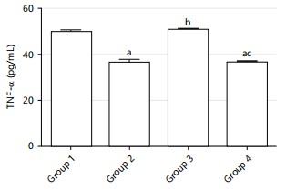
Influence of flavonoid-rich cocoa aqueous extract on TNF-α and IL-6 activities: The anti-inflammatory studies indicate a substantial reduction in TNF-α activity (p<0.05) in group 2 and 4 when compared to the control group (CONT) and group 3. However, TNF-α activity in group 3 did not show a significant difference (p<0.05) when compared to the CONT group. Moreover, TNF-α activity in group 3 was marginally higher, though not significantly, compared to group 2 and 4 (p<0.05). These results were illustrated in Fig. 5, suggest that the cocoa flavonoid aqueous extract has a more pronounced anti-inflammatory effect in group 2 and 4.
Similarly, IL-6 activity was significantly decreased (p<0.05) in group 2 and 4 compared to both the CONT group and group 3. In contrast, IL-6 activity in group 3 was not significantly different (p<0.05) from the CONT group. Additionally, IL-6 activity in group 3 was slightly higher, but not significantly so, compared to group 2 and 4 (p<0.05). These findings, as depicted in Fig. 6, further demonstrated that the cocoa flavonoid aqueous extract effectively reduces inflammation in group 2 and 4.

|
|
|
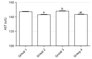
Influence of flavonoid-rich cocoa aqueous extract on AST, ALT, ALP, albumin and total protein: The liver function marker results indicate a notable reduction in AST activity (p<0.05) in group 2 and 4 when compared to the control group (CONT) and group 3. In group 3, AST activity was not significantly different (p<0.05) from the CONT group but was slightly higher, though not significantly, compared to group 2 and 4 (p<0.05). These findings, depicted in Fig. 7, suggest that the cocoa flavonoid aqueous extract may have a protective effect on liver function, particularly in group 2 and 4.
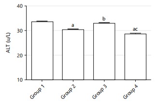
Similarly, ALT activity showed a significant decrease (p<0.05) in group 2 and 4 compared to both the CONT group and group 3. In contrast, ALT activity in group 3 was not significantly different (p>0.05) from the CONT group but was insignificantly higher compared to group 2 and 4 (p<0.05). This trend is further confirmed by another set of results showing a significant decrease in ALT activity (p<0.05) in group 2 and 4 compared to CONT and group 3, while group 3 exhibited significantly increased ALT activity (p<0.05) compared to CONT. Additionally, ALT activity in group 3 was significantly higher (p<0.05) than in group 2 and 4, as shown in Fig. 8 and 9. These results imply that the cocoa flavonoid aqueous extract effectively reduces liver inflammation and damage in group 2 and 4.
|

|

|
Albumin levels in group 2 and 4 were not significantly different (p<0.05) compared to the CONT group, while levels were significantly increased (p<0.05) in group 3, 2 and 4. This indicates that the cocoa flavonoid aqueous extract does not adversely affect albumin levels, as illustrated in Fig. 10. Additionally, there was no significant difference (p<0.05) in total protein levels in group 2 and 4 compared to CONT, while the levels were significantly increased (p<0.05) in group 3, 2 and 4. This is depicted in Fig. 11 and suggests that the extract maintains protein synthesis and overall liver function stability.
DISCUSSION
In this study, GSH, SOD, CAT and MDA were evaluated as indices of oxidative status.
The results of this study showed that rats fed cocoa flavonoids had significantly higher levels of endogenous antioxidants such as superoxide dismutase (SOD) and catalase and decreased levels of lipid peroxidation products. These findings suggest that cocoa flavonoids improve antioxidant capacity and help alleviate high-fat diet-related oxidative stress in Wistar rats. Known for its high flavonoid content, cocoa is a promising source of natural antioxidants. Flavonoids such as epicatechin and catechin can scavenge free radicals, thus preventing oxidative stress caused by a high-fat diet20,21. The antioxidant potential of cocoa flavonoids is further supported by their ability to enhance the activity of endogenous antioxidant enzymes, including superoxide dismutase (SOD) and catalase20,21. Oxidative stress, characterized by an imbalance between reactive oxygen species (ROS) production and antioxidant defense, is a hallmark of obesity and related metabolic disorders. These findings were consistent with the broader literature and highlighted the role of flavonoids in enhancing cellular defenses against oxidative damage22. The results showed increased activity of SOD, GSH and CAT in lean and obese rats indicating an effective oxidative state. The antioxidant properties of cocoa flavonoids in this study agreed with a study by Katz et al.21. In Wistar rats fed a high-fat diet, administration of flavonoid-rich cocoa extract may alleviate oxidative stress by neutralizing reactive oxygen species (ROS) and preventing lipid peroxidation. Such antioxidants can extend beyond the cellular barrier, affecting systemic signs of oxidative stress and contributing to the maintenance of redox homeostasis. Recent studies have further corroborated these findings, demonstrating the protective effects of cocoa flavonoids23.
Chronic inflammation is a hallmark of obesity and related metabolic disorders. A high-fat diet triggers the release of pro-inflammatory cytokines, which promote low-grade inflammation. Cocoa flavonoids have shown anti-inflammatory properties by modulating key cytokines and inflammatory mediators24. The potential of cocoa polyphenols to attenuate inflammatory responses is particularly important in the case of fatty food-induced inflammation. The anti-inflammatory effect of cocoa is due to its ability to inhibit the expression of cytokines such as tumor necrosis factor-alpha (TNF-α) and interleukin-6 (IL-6)24. These cytokines play a central role in the initiation and propagation of inflammation and their suppression by cocoa flavonoids suggests a plausible mechanism for dampening the inflammatory cascade induced by a high-fat diet. In an experimental setting, the administration of flavonoid-rich cocoa extract to Wistar rats fed a high-fat diet is thought to suppress the inflammatory environment, alleviating the burden of chronic inflammation associated with obesity-related complications. In this study, the aqueous extract of flavonoid-rich cocoa showed promising effects, which were observed in treated rats with a significant decrease in serum levels of TNF-α and IL-6, indicative of the anti-inflammatory potential of the flavonoid-rich extract of cocoa. Newer research supports these findings, showing that cocoa flavonoids can effectively reduce inflammation markers in metabolic disorders25.
The liver, as the central organ of energy metabolism, is prone to metabolic disorders caused by fatty foods. Increased liver enzyme markers such as alanine transaminase (ALT) and aspartate transaminase (AST) are signs of liver dysfunction and are often seen in fatty liver and inflammation. Previous studies have shown that cocoa flavonoids can protect against diet-induced liver damage. A reduction in liver enzymes and amelioration of liver steatosis in mice fed a high-fat diet after supplementation with cocoa
flavonoids were reported by Lee et al.23. This suggests that cocoa can have a hepatoprotective effect, which can occur through the modulation of lipid metabolism and the weakening of inflammatory processes in the liver and this suggests that Wistar rats fed a high-fat diet, administration of an aqueous extract containing flavonoid-rich cocoa had a positive effect on liver enzyme markers, reflecting liver health. Serum proteins, including the predominant protein albumin, play an important role in oxidative stress and liver disease. In liver diseases, oxidative stress is often increased due to various factors such as inflammation and impaired antioxidant defenses26.
Albumin, an important serum protein, is not only synthesized by the liver but also acts as an important antioxidant. It is recognized for its ability to scavenge free radicals and protect against oxidative damage. However, in liver diseases, especially when the liver is damaged, albumin synthesis and antioxidant capacity can be impaired, increasing susceptibility to oxidative stress27. This finding revealed rats exposed to cocoa flavonoid-rich extract displayed increased albumin levels, which is suggestive of the protective role of the extract on liver dysfunction caused by the high-fat diet. Studies have highlighted an association between decreased serum albumin levels and the severity of liver disease, indicating its potential role as a biomarker of liver dysfunction. In addition, low albumin levels have been associated with signs of increased oxidative stress in liver disease28. Serum proteins, including albumin, are essential for maintaining physiological homeostasis and deviations from normal levels may indicate metabolic imbalance. In agreement with the previous findings, results from this investigation suggest that rats fed cocoa flavonoid aqueous extract had significant preservation of serum protein and albumin compared with a high-fat diet alone29.
The discovery that flavonoid-rich cocoa extract reduces oxidative stress, inflammation and liver dysfunction caused by high-fat diets in Wistar rats has significant applications and implications. This suggests that cocoa extract could be used as a dietary supplement or functional food ingredient to treat metabolic illnesses associated with high-fat diets, such as Nonalcoholic Fatty Liver Disease (NAFLD). Cocoa flavonoids' anti-inflammatory and antioxidant activities may play an important role in preventing or treating chronic disorders linked to oxidative stress and inflammation. Future studies should focus on determining the precise molecular pathways by which cocoa flavonoids exert their protective effects, as well as performing clinical trials to assess their efficacy and safety in people. Furthermore, investigating alternative formulations and dosages may improve the therapeutic potential of cocoa extracts in the setting of diet-induced metabolic diseases.
CONCLUSION
This study explored the impact of flavonoid-rich cocoa aqueous extract on antioxidant, anti-inflammatory and liver enzyme markers in high-fat diet Wistar rats and presented a promising research avenue. The antioxidant and anti-inflammatory characteristics of cocoa flavonoids position them as potential therapeutic agents for mitigating the harmful effects of a high-fat diet. As research advances, cocoa could emerge as a valuable dietary supplement in the multifaceted treatment of obesity and associated metabolic disorders.
SIGNIFICANCE STATEMENT
This work is important because it may identify cocoa flavonoids as a natural defense against inflammation and oxidative stress brought on by a high-fat diet. The study highlights the potential of cocoa extract to enhance metabolic health and liver function by exhibiting its hepatoprotective and anti-inflammatory benefits in Wistar rats given a high-fat diet. This could open the door for the creation of nutraceuticals or functional foods that are intended to lessen the negative health consequences of high-fat diets, improving public health and aiding in the management and prevention of metabolic disorders.
ACKNOWLEDGMENT
The authors would like to acknowledge Alhaji Musa Adamu for his support throughout this work.
REFERENCES
- Ling, C. and T. Rönn, 2019. Epigenetics in human obesity and type 2 diabetes. Cell Metab., 29: 1028-1044.
- Abdisa, K.B., E. Szerdahelyi, M.A. Molnár, L. Friedrich and Z. Lakner et al., 2024. Metabolic syndrome and biotherapeutic activity of dairy (cow and buffalo) milk proteins and peptides: Fast food-induced obesity perspective-A narrative review. Biomolecules, 14.
- Goya, L., M.Á. Martín, B. Sarriá, S. Ramos, R. Mateos and L. Bravo, 2016. Effect of cocoa and its flavonoids on biomarkers of inflammation: Studies of cell culture, animals and humans. Nutrients, 8.
- Davison, K., A.M. Coates, J.D. Buckley and P.R.C. Howe, 2008. Effect of cocoa flavanols and exercise on cardiometabolic risk factors in overweight and obese subjects. Int. J. Obesity, 32: 1289-1296.
- Lee, K.W., Y.J. Kim, H.J. Lee and C.Y. Lee, 2003. Cocoa has more phenolic phytochemicals and a higher antioxidant capacity than teas and red wine. J. Agric. Food Chem., 51: 7292-7295.
- Caminiti, R., C. Carresi, R. Mollace, R. Macrì and F. Scarano et al., 2024. The potential effect of natural antioxidants on endothelial dysfunction associated with arterial hypertension. Front. Cardiovasc. Med., 11.
- Grassi, D., S. Necozione, C. Lippi, G. Croce and L. Valeri et al., 2005. Cocoa reduces blood pressure and insulin resistance and improves endothelium-dependent vasodilation in hypertensives. Hypertension, 46: 398-405.
- Mainini, G., S. Ercolano, R. de Simone, I. Iavarone, R. Lizza and M. Passaro, 2024. Dietary supplementation of myo-inositol, cocoa polyphenols, and soy isoflavones improves vasomotor symptoms and metabolic profile in menopausal women with metabolic syndrome: A retrospective clinical study. Medicina, 60.
- Helmut, S., S. Tankred, H. Christian and K. Malte, 2005. Cocoa polyphenols and inflammatory mediators. Am. J. Clin. Nutr., 81: 304S-312S.
- Asiedu-Gyekye, I.J., T.G. Borovskaya, M.E. Poluektova, А.V. Vychuzhanina and Y.А. Shchemerovа et al., 2021. Reproductive toxicity of Theobroma cacao: Increase in survival index, nongenotoxic, and proimplantation potential. J. Toxicol., 2021.
- Igbayilola, Y.D. and M.G. Gujja, 2024. Alpha-amylase and alpha-glucosidase upregulated glucose homeostasis in high-fat fed Wistar rats supplemented with cocoa flavonoid-rich aqueous. Food Biosci., 59.
- Bag, G.C., P.G. Devi, and T. Bhaigyabati, 2015. Assessment of total flavonoid content and antioxidant activity of methanolic rhizome extract of three hedychium species of Manipur Valley. Int. J. Pharm. Sci. Rev. Res., 30: 154-159.
- Senguttuvan, J., S. Paulsamy and K. Karthika, 2014. Phytochemical analysis and evaluation of leaf and root parts of the medicinal herb, Hypochaeris radicata L. for in vitro antioxidant activities. Asian Pac. J. Trop. Biomed., 4: S359-S367.
- Makkar, H.P.S., P. Siddhuraju and K. Becker, 2007. Saponins. In: Plant Secondary Metabolites, Makkar, H.P.S., P. Siddhuraju and K. Becker (Eds.), Humana Press, Totowa, New Jersey, ISBN: 978-1-58829-993-2, pp: 93-100.
- Benedict, S.R., 1909. A reagent for the detection of reducing sugars. J. Biol. Chem., 5: 485-487.
- Harborne, J.B., 1973. Phytochemical Methods: A Guide to Modern Techniques of Plant Analysis. Chapman & Hall, Boundary Row, London, ISBN: 9780412105401, Pages: 278.
- Amin, K.A. and M.A. Nagy, 2009. Effect of carnitine and herbal mixture extract on obesity induced by high fat diet in rats. Diabetol. Metab. Syndr., 1.
- Vilela, D.D., L.G. Peixoto, R.R. Teixeira, N.B. Baptista and D.C. Caixeta et al., 2016. The role of metformin in controlling oxidative stress in muscle of diabetic rats. Oxid. Med. Cell. Longevity, 2016.
- Umukoro, S., H.A. Kalejaye, B. Ben-Azu and A.M. Ajayi, 2018. Naringenin attenuates behavioral derangements induced by social defeat stress in mice via inhibition of acetylcholinesterase activity, oxidative stress and release of pro-inflammatory cytokines. Biomed. Pharmacother., 105: 714-723.
- Uchiyama, M. and M. Mihara, 1978. Determination of malonaldehyde precursor in tissues by thiobarbituric acid test. Anal. Biochem., 86: 271-278.
- Katz, D.L., K. Doughty and A. Ali, 2011. Cocoa and chocolate in human health and disease. Antioxid. Redox Signaling, 15: 2779-2811.
- Ding, W., X. Yang, K. Lai, Y. Jiang and Y. Liu, 2024. The potential of therapeutic strategies targeting mitochondrial biogenesis for the treatment of insulin resistance and type 2 diabetes mellitus. Arch. Pharm. Res., 47: 219-248.
- Lee, H., K. Colin, A. Asmaa, A.K. Paul, C.S. Jeffrey, B.R. Eric and C. Aedín, 2012. Effects of chocolate, cocoa, and flavan-3-ols on cardiovascular health: A systematic review and meta-analysis of randomized trials. Am. J. Clin. Nutr., 95: 740-751.
- Park, N.W., E.S. Lee, K.B. Ha, S.H. Jo, H.M. Kim, M.H. Kwon and C.H. Chung, 2023. Umbelliferone ameliorates hepatic steatosis and lipid-induced ER stress in high-fat diet-induced obese mice. Yonsei Med. J., 64: 243-250.
- Selmi, C., C.A. Cocchi, M. Lanfredini, C.L. Keen and M.E. Gershwin, 2008. Chocolate at heart: The anti-inflammatory impact of cocoa flavanols. Mol. Nutr. Food Res., 52: 1340-1348.
- Rondanelli, M., M.A. Faliva, A. Miccono, M. Naso and M. Nichetti et al., 2018. Food pyramid for subjects with chronic pain: Foods and dietary constituents as anti-inflammatory and antioxidant agents. Nutr. Res. Rev., 31: 131-151.
- Halliwell, B. and J.M.C. Gutteridge, 2015. Free Radicals In Biology And Medicine. 5th Edn., Oxford University Press, Oxford, England, ISBN: 9780198717485, Pages: 905.
- Oettl, K. and R.E. Stauber, 2007. Physiological and pathological changes in the redox state of human serum albumin critically influence its binding properties. Br. J. Pharmacol., 151: 580-590.
- Poli, G. and M. Parola, 1997. Oxidative damage and fibrogenesis. Free Radical Biol. Med., 22: 287-305.
How to Cite this paper?
APA-7 Style
Dimeji,
I.Y., Gujja,
G.M., Adeoye,
S.W., Jabba,
H.L., ,
J.S., Baba,
Z.M. (2024). Flavonoid-Rich Cocoa Extract Alleviates Oxidative Stress, Inflammation and Liver Dysfunction Induced by High-Fat Diets in Wistar Rats. Research Journal of Phytochemistry, 18(1), 20-33. https://doi.org/10.3923/rjp.2024.20.33
ACS Style
Dimeji,
I.Y.; Gujja,
G.M.; Adeoye,
S.W.; Jabba,
H.L.; ,
J.S.; Baba,
Z.M. Flavonoid-Rich Cocoa Extract Alleviates Oxidative Stress, Inflammation and Liver Dysfunction Induced by High-Fat Diets in Wistar Rats. Res. J. Phytochem 2024, 18, 20-33. https://doi.org/10.3923/rjp.2024.20.33
AMA Style
Dimeji
IY, Gujja
GM, Adeoye
SW, Jabba
HL,
JS, Baba
ZM. Flavonoid-Rich Cocoa Extract Alleviates Oxidative Stress, Inflammation and Liver Dysfunction Induced by High-Fat Diets in Wistar Rats. Research Journal of Phytochemistry. 2024; 18(1): 20-33. https://doi.org/10.3923/rjp.2024.20.33
Chicago/Turabian Style
Dimeji, Igbayilola, Yusuff, Grema Mariam Gujja, Saka Waidi Adeoye, Hamidu, Lawan Jabba, Jibrin, Sherifdeen , and Zakari Muhammed Baba.
2024. "Flavonoid-Rich Cocoa Extract Alleviates Oxidative Stress, Inflammation and Liver Dysfunction Induced by High-Fat Diets in Wistar Rats" Research Journal of Phytochemistry 18, no. 1: 20-33. https://doi.org/10.3923/rjp.2024.20.33

This work is licensed under a Creative Commons Attribution 4.0 International License.