
Phytochemical Characterization and Antibacterial Activity of Lantana camara L. Seed Extracts
| Received 09 Mar, 2025 |
Accepted 20 Sep, 2026 |
Published 30 Sep, 2026 |
Background and Objective: Lantana camara L. is a medicinal plant rich in bioactive phytochemicals traditionally used for treating infections and various ailments. The main objective was to characterize the bioactive compounds present in Lantana camara seeds and to evaluate their antioxidant and antibacterial activities. Materials and Methods: Two solvents were used for extraction from 100 g of seed powder: Pure ethanol and a hydroethanolic mixture containing 70% water and 30% ethanol. The extracts were the subjected to phytochemical screening, quantitative analysis of phenolic compounds, flavonoids, and tannins, as well as antioxidant activity assays using DPPH and ABTS, and antibacterial susceptibility tests. Data were analyzed in GraphPadPrism 8, with results as Mean±SD at p≤0.05. Results: The hydroethanolic mixture produced a higher extraction yield (10.30%) compared to pure ethanol (4.736%). However, the ethanolic extract was richer in phenolic compounds (67.10 mg gallic acid equivalents per gram of dry extract), flavonoids (354.83 mg quercetin equivalents per gram of dry extract), and tannins (234.50 mg catechin equivalents per gram of dry extract). It also showed stronger antioxidant activity, with an IC50 of 2942.83 μg/mL for DPPH and 2100.27 μg/mL for ABTS, compared to the hydroethanolic extract, which had IC50 values of 3173.84 μg/mL for DPPH and 2832.71 μg/mL for ABTS, although both were less effective than quercetin. Finally, only the ethanolic extract demonstrated significant antibacterial activity against all six tested strains, including Pseudomonas aeruginosa and Klebsiella pneumoniae. Conclusion: Ethanol appears to be the most effective solvent for extracting bioactive compounds from Lantana camara seeds, due to its ability to concentrate the secondary metabolites responsible for antioxidant and antibacterial activities. These findings confirm the pharmacological potential of this plant and support its use in traditional medicine.
INTRODUCTION
Plants are an essential resource for sustainable human development due to their richness in bioactive compounds with well-recognized therapeutic properties1. Among them, certain species stand out for their particularly interesting medicinal potential.
One such species is Lantana camara L., a hardy, evergreen shrub recognizable by its characteristic odor. It grows between 0.5 and 3 m tall and may or may not bear small thorns on its branches. Its leaves, when crushed, release a strong smell. Flowering generally occurs between June and October, with flowers displaying various colors: Yellow, white, pink, or red. The fruits, which turn black when ripe, are toxic in their unripe state2.
Originally native to Central and South America, Lantana camara has become widely naturalized in many tropical regions, particularly in Africa. Today, it is cultivated in several tropical countries, often in pots or planters3.
From a medicinal standpoint, the Lantana genus is widely used in traditional medicine. Its essential oil is known for its antibacterial, antifungal, and repellent properties4. The leaves and flowers are commonly used to repel insects.
Numerous studies have highlighted the biological activities of Lantana camara extracts. For example, ethanolic extracts of the leaves have shown antioxidant and antibacterial activity, as reported by Reddy, Patil, and Kumbhar2,5. Methanolic extracts have demonstrated antidiabetic, antiproliferative, and anti-ulcer properties4,5. Gidwani et al.6 showed that aqueous leaf extracts of Lantana camara possess anti-inflammatory and analgesic effects.
With regard to the essential oil, antimicrobial activity has been observed, with caryophyllene oxide (23.015%) and spathulenol (13.421%) identified as the major components7. Antifungal properties have also been reported8, with the main constituents identified as (E)-β-caryophyllene (40.8%) and α-humulene (21.2%)9.
Furthermore, in Togo, the insecticidal activity of Lantana camara essential oil has been demonstrated10.
However, the seeds of this plant have not yet been thoroughly investigated, despite their valuable potential that can be used as medicinal compounds, minerals, and nutrients. In order to fully exploit their biological potential, it is essential to better understand their chemical composition. Therefore, the purpose of this study is to evaluate the phytochemical composition and biological activities of Lantana camara seed extracts, in order to better assess their potential health benefits.
MATERIALS AND METHODS
Study area and duration: This study was conducted in Lomé, in the Golfe prefecture of Togo. Seeds of Lantana camara L. were collected at the University of Lomé, an area characterized by a sub-equatorial or Guinean climate (humid tropical climate) influenced by its coastal location, as well as by diverse vegetation. The experimental work, including sample collection, extract preparation, phytochemical analyses, and antibacterial activity tests, was carried out over a twelve-month period, from June 2024 to May 2025.
Collected plant specimens: Lantana camara seeds were harvested in Lomé, situated in the Maritime Region of Togo. The plant was taxonomically identified at the Laboratory of Botany and Plant Ecology, within the Department of Botany, Faculty of Science, University of Lomé, and the specimen was catalogued under the reference number TOGO15902. Following collection, the seeds were air-dried at ambient temperature (approximately 27°C) for a period of 14 days. After drying, they were finely ground using an electric grinder to produce a uniform powder.
Preparation of extracts: For the extraction procedure, 100 g of powdered seeds were subjected to maceration in 1 L of ethanol to obtain the ethanolic extract, and in 1 L of a 70:30 ethanol-distilled water mixture for the hydroethanolic extract. The maceration was carried out over 72 hrs with occasional stirring to enhance solvent penetration. After this period, each mixture was filtered using Whatman filter paper. The resulting filtrates were concentrated at 40°C using a rotary evaporator (BUCHI model) to recover the respective dry extracts.
Phytochemical profiling: Phytochemical profiling was conducted on both extracts of Lantana camara seeds. Extractions and characterization tests were performed using standard methods based on colorimetric and precipitation reactions11-13. The presence of anthraquinones, flavonoids, anthocyanins, alkaloids, saponins, tannins, terpenes, and terpenoids was investigated. An analysis was performed to detect the presence of various phytochemical groups, including anthraquinones, flavonoids, anthocyanins, alkaloids, saponins, tannins, as well as terpenes and terpenoids.
Tannin detection: To test for tannins, 1 mL of the plant extract was combined with a few drops of 1% ferric chloride (FeCl3) solution. The formation of a dark blue, green, or black coloration was taken as indicative of the presence of tannins.
Flavonoid detection: For the identification of flavonoids, 5 mL of the extract were mixed with an equal volume of alcoholic hydrochloric acid (Shinoda reagent), followed by the addition of a small quantity of magnesium powder. The development of an orange hue indicated the presence of flavones, red signified flavonols, and violet suggested flavanones.
Alkaloid detection: To detect alkaloids, 1 g of the extract was macerated in 25 mL of 5% hydrochloric acid for 24 hrs. After filtration, the treatment of 1 mL of the filtrate was made with 5 drops of Mayer’s reagent. The appearance of a pale yellow or cloudy precipitate confirmed the presence of alkaloids.
Terpenoids detection: The detection of terpenoids was performed using the Liebermann-Burchard reagent. Approximately 2 mL of plant extract were placed in a test tube, followed by the addition of 2 mL of chloroform. The mixture was shaken to ensure proper homogenization. Then, 3 mL of concentrated sulfuric acid were carefully added along the inner wall of the tube using a pipette, forming a distinct layer beneath the mixture. The test tube was allowed to stand undisturbed to facilitate the development of a distinct interface between the two liquid phases. The appearance of a reddish, brownish, or violet coloration at the junction of the layers was considered an indicator of terpenoid presence in the extract.
Mucilages detection: To perform the test, 1 mL of a 10% diluted extract was placed into a test tube, and 5 mL of absolute ethanol was then added. The formation of a flocculent precipitate within approximately ten minutes suggests the presence of mucilage compounds.
Saponins detection: Saponins were detected by the foam index method, which is based on the extent of dilution of an aqueous decoction of the plant that produces a persistent foam under specific conditions. A measured volume of the plant extract was diluted to a final volume of 100 mL, followed by serial 1:10 dilutions into 10 separate test tubes. Each tube was then shaken vertically 30 times over a 15 sec period. After being left to stand undisturbed for 15 min, the height of the resulting foam was recorded. If it exceeded 1 cm in any of the tubes, the corresponding dilution was considered as the foam index.
Test for terpenes and sterols: A quantity of 0.5 g of the extract was mixed with 10 mL of ethanol preheated to 70°C and agitated for 30 min. Subsequently, 10 mL of distilled water and 2 mL of a 10% (v/v) lead acetate solution were added. After allowing the mixture to stand for 15 min, 2 mL of a 10% aqueous disodium phosphate solution was added to the filtrate. Following another 15 min resting period, the resulting solution was transferred into a separating funnel and subjected to triple extraction using 5 mL portions of chloroform. The chloroform layers were combined, dried over anhydrous sodium sulfate, and concentrated by evaporation. The dry residue was then dissolved again in a small volume of acetic acid, followed by the addition of 3 mL of a reagent mixture containing acetic anhydride and sulfuric acid. The emergence of a violet, blue, or green coloration was taken as a positive indication of triterpenoids.
Anthocyanins detection: An aliquot of 1 mL of the extract was initially acidified with several drops of 5% hydrochloric acid, followed by basification through the gradual addition of diluted ammonia. A deepening of the red color, turning to bluish-purple or green, indicates the presence of anthocyanins.
Determination of total phenolic content: The total phenolic content of the different extracts and their respective fractions was quantitatively determined using the Folin-Ciocalteu colorimetric assay14. Briefly, 200 μL of each extract (prepared at a concentration of 1 mg/mL) was combined with 1.5 mL of Folin-Ciocalteu reagent previously diluted 1:10 with distilled water. After an incubation period of 5 min at room temperature (25°C), 1.5 mL of a Sodium Carbonate (Na2CO3) solution at a concentration of 70 g/L were added to the mixture. The reaction was allowed to proceed in the dark for 90 min. Absorbance was then measured at 760 nm using a UV-VIS spectrophotometer (model 5100B), with the appropriate blank used for calibration. Gallic acid, in concentrations ranging from 0 to 500 μg/mL, served as the standard for the calibration curve. Results were expressed as milligrams of gallic acid equivalents per gram of dry extract (mg GAE/g dry extract), calculated by interpolation from the standard curve equation: = 0.0139+0.0046 (avec R2 = 0.9924) obtained from the gallic acid standard solutions. Each measurement was performed in triplicate for each extract.
Quantitative analysis of flavonoid: To estimate the total flavonoid content in the extracts, a colorimetric method based on Aluminum Chloride (AlCl3) was employed, following previously established protocols15,16. An aliquot of 300 μL of the ethanolic extract was first diluted with 1.2 mL of distilled water. Subsequently, 90 μL of a 5% Sodium Nitrite (NaNO2) solution was added. After incubating the mixture for 5 min at room temperature, 60 μL of a 10% aluminum chloride solution was introduced. This was followed by the addition of 600 μL of 1 M Sodium Carbonate (Na2CO3) and 750 μL of distilled water. The mixture was then left to stand for another 5 min. After vortexes, the absorbance was recorded at 510 nm using a UV-Visible spectrophotometer (model 5100B), with a blank used as the reference. The flavonoid concentration was determined from a standard curve constructed using quercetin solutions ranging from 0 to 500 μg/mL. The resulting standard curve equation was: y = 0.0006x-0.0089 (R2 = 0.9773) where R2 represents the coefficient of determination. The results were reported as milligrams of quercetin equivalents per gram of dry extract (mg QE/g DE).
Quantitative analysis of tannins: The vanillin method in the presence of HCl, based on the reaction of condensed tannins with vanillin in an acidic medium to form red-colored complexes, was used for their determination17,18. The condensed tannin content was determined using the vanillin assay. A volume of 50 μL of each extract was added to 1500 μL of 4% vanillin/ethanol solution, then mixed thoroughly. Subsequently, 750 μL of concentrated hydrochloric acid was added. The resulting mixture was allowed to react at room temperature for 20 min. The absorbance was recorded at 550 nm against a reagent blank using a UV-VIS 5100B spectrophotometer. The concentration of condensed tannins was expressed as milligrams of catechin equivalents per gram of dry extract (mg CE/g DE). Quantification was based on a calibration curve constructed using catechin standard solutions in the range of 017 to 1000 μg/mL, with the corresponding regression equation: y = 0.0002x+0.0036 (R2 = 0.9856).
Evaluation of antioxidant properties of Lantana camara extracts in vitro
DPPH (2,2-Diphenyl-1-picrylhydrazyl) assay: The DPPH (2,2-diphensyl-1-picrylhydrazyl) assay is commonly employed for the rapid and simple assessment of antioxidant activity, owing to the compound’s stability as a free radical and the ease of measurement. The DPPH exhibits a strong absorbance at 517 nm in the visible spectrum. In this study, the evaluation of DPPH radical scavenging activity was conducted based on the methodology outlined by Evenamede et al.19.
Assessment of antioxidant activity using ABTS radical: The antioxidant potential of the plant extracts was assessed through the ABTS· (2,2 -azino-bis(3-ethylbenzothiazoline-6-sulfonic acid)) radical cation decolorization assay20. The ABTS· radical was produced by mixing equal volumes of an aqueous ABTS solution (7 mM) and potassium persulfate (2.45 mM). This mixture was kept in the dark at room temperature for 12 to 16 hrs to allow radical formation. Before the assay, the resulting solution was diluted with ethanol until the absorbance reached approximately 0.700 at 734 nm. For each test, 50 μL of the plant extract was combined with 1.95 mL of the diluted ABTS· solution, and the absorbance was recorded after 30 min. Corresponding blanks containing only the solvent were included in all assays. Each measurement was carried out in triplicate to ensure reproducibility. The percentage of inhibition at 734 nm was determined using the following equation:
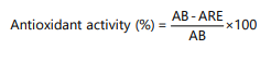 |
Where:
| AB | = | Absorbance of the ABTS solution mixed with ethanol (control) | |
| AE | = | Absorbance of the ABTS solution mixed with the test extract or standard |
Quercetin was used as the reference compound21.
Antibacterial test22
Collection of microorganisms: The bacterial strains used were:
| • | Gram-negative bacteria: Escherichia coli ATCC 25922, Klebsiella pneumoniae ATCC 700603, Pseudomonas aeruginosa ATCC 27853, Salmonella typhimurium ATCC 14028, and Shigella flexneri ATCC 12022 | |
| • | Gram-positive bacterium: Staphylococcus aureus ATCC 29213. These reference bacterial strains were obtained from the National Institute of Hygiene (INH) in Togo, Bacteriology Laboratory |
Sensitivity test of the extracts: Bacterial inocula were prepared by suspending 24 hrs old colonies in 10 mL of sterile physiological saline. The suspension’s turbidity was adjusted to match 0.5 on the McFarland scale using a densitometer. Antibacterial activity was evaluated through the agar well diffusion technique. Mueller-Hinton agar plates were uniformly seeded with the standardized bacterial suspension. Wells were aseptically created in the agar and each was loaded with 50 μL of plant extract at a concentration of 100 mg/mL. The plates were incubated at 37°C for 24 hrs. Zones of inhibition surrounding the wells were measured with a caliper. All experiments were conducted in triplicate. The antibacterial efficacy of the extracts was interpreted based on the classification system proposed by Ponce et al.23. Gentamicin was used as the antibiotic for the positive control.
Measurement of the minimum inhibitory and bactericidal concentrations: The MIC and MBC were assessed using the macrodilution technique in liquid medium, carried out in sterile hemolysis tubes24,25. A stock solution of the plant extract (100 mg/mL) was used to prepare a series of two-fold dilutions in Mueller-Hinton broth, yielding final concentrations ranging from 100 to 1.56 mg/mL. For each test, 1 mL of the diluted extract was combined with 1 mL of bacterial suspension (105 CFU/mL), previously standardized to 0.5 McFarland turbidity:
| • | Growth control: Mueller-Hinton broth+inoculum (without extract) | |
| • | Sterility control: Mueller-Hinton broth only (without inoculum) |
The tubes were wrapped in aluminum foil and kept at 37°C for 24 hrs. The minimum inhibitory concentration (MIC) was determined as the lowest extract concentration that prevented any visible cloudiness, indicating suppression of bacterial growth.
The minimum bactericidal concentration (MBC) refers to the lowest concentration of the extract that results in a 99.99% reduction in bacterial viability, meaning less than 0.01% of the initial bacterial population remains after 24 hrs of incubation.
For MBC determination:
| • | Serial dilutions of the initial inoculum (from 10–1 to 10–4) were prepared | |
| • | Using a calibrated loop (2 μL), these dilutions were inoculated onto Mueller-Hinton agar (MHA) plates (5 cm streaks) | |
| • | After incubation at 37°C for 24 hrs, the plates (series B) were compared to those used for MIC determination (series A) | |
| • | The MBC corresponds to the lowest concentration where bacterial growth is less than 0.01% compared to the growth control |
The MBC/MIC ratio allows determination of the extract’s mode of action26:
| • | If the MBC to MIC ratio is less than or equal to 2, the extract is considered bactericidal | |
| • | If the MBC to MIC ratio is greater than 2, the extract is classified as bacteriostatic |
Data analysis: Data analysis was performed using GraphPad Prism version 8, which also served to generate graphs and histograms. Microbiological results were evaluated to compare the effectiveness of extracts from mature Lantana camara seeds against the various tested bacterial strains. The results are presented as mean values with standard deviation (±SD), and statistical significance was defined at a threshold of p≤0.05.
RESULTS AND DISCUSSION
Extraction yield: In this study, two different solvents were used to extract bioactive compounds from 100 g of powdered ripe seeds of Lantana camara: Ethanol and a hydroethanolic mixture (70% water/30% ethanol). The extraction yields are reported in Table 1. The results indicate that the hydroethanolic solvent produced a higher extraction yield (10.30%) with a dry extract weight of 10.30 g compared to ethanol extraction, which yielded 4.736% with a dry extract weight of 4.736 g.
| Table 1: | Extraction yields according to the solvent used | |||
| Condition | Solvent | Dry extract | Yield (%) |
| Condition 1 | Ethanol | 4.736 g | 4.74% |
| Condition 2 | Hydroethanol | 10.30 g | 10.30% |
| Table 2: | Phytochemical screening results of Lantana camara extracts | |||
| Phytochemicals searched | Ethanolic extract | Hydroethanolic extract |
| Alkaloids | - | - |
| Flavonoids | + | + |
| Tannins | + | + |
| Saponins | - | + |
| Reducing sugars | - | + |
| Anthocyanins | - | - |
| Terpenes and sterols | + | + |
| Mucilages | - | - |
| +: Present and =: Absent or low concentration | ||
|
|
Analysis of phytochemical constituents in various extracts: Phytochemical analysis of the ethanolic and hydroethanolic (30/70) extracts of Lantana camara demonstrated the presence of various bioactive compound groups. Both extracts contained flavonoids, tannins, reducing sugars, and terpenes/sterols. However, alkaloids, anthocyanins, and mucilages were either undetectable or found in trace amounts. Notably, saponins were identified exclusively in the hydroethanolic extract (Table 2).
Quantitative test results
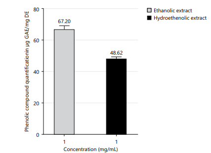
Evaluation of total phenolic content: The total polyphenol content varied between 48.62±1.59 and 67.10±2.22 mg gallic acid equivalents per gram of dry extract (mg GAE/g DE) for the hydroethanolic (70/30) and ethanolic extracts, respectively, measured at a concentration of 1 mg/mL (Fig. 1).
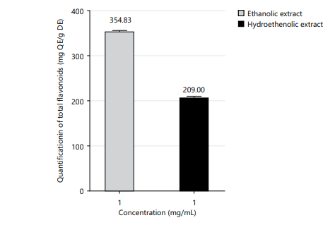
Determination of total flavonoid content: The total flavonoid levels varied between 209.00±3.53 and 354.83±3.06 mg quercetin equivalents per gram of dry extract (mg QE/g DE) for the hydroethanolic (70/30) and ethanolic extracts, respectively, measured at a concentration of 1 mg/mL (Fig. 2).
|
|
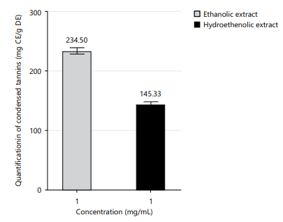
Quantification of condensed tannins: The condensed tannin levels were found to range from 145.33±3.68 to 234.50±5.30 mg catechin equivalents per gram of dry extract (mg CE/g DE) in the hydroethanolic (70/30) and ethanolic extracts, respectively, at a concentration of 1 mg/mL (Fig. 3).
Evaluation of antioxidant activity
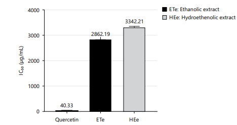
DPPH radical scavenging assay: The DPPH assay (Fig. 4) revealed a significantly higher antioxidant activity for quercetin (IC50 = 40 μg/mL) compared to the plant extracts. The ethanolic extract exhibited a better free radical scavenging capacity (IC50 = 2862 μg/mL) than the hydroethanolic extract (IC50 = 3342 μg/mL), although both showed markedly lower activity than quercetin.
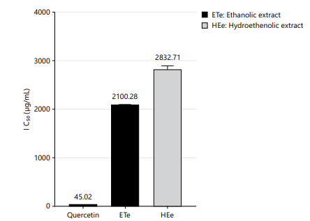
Determination of antioxidant capacity of extracts through the ABTS· assay: The findings of the ABTS assay (Fig. 5) indicate that quercetin (IC50 = 45.02 μg/mL) exhibits significantly higher radical-scavenging activity compared to the seed extracts of Lantana camara, as evidenced by its low IC50 values. Among the tested extracts, the ethanolic extract demonstrated greater antioxidant capacity (IC50 = 2100.27 μg/mL) than the hydroethanolic extract (IC50 = 2832.71 μg/mL), although both were markedly less active than quercetin.
|
| Table 3: | Antimicrobial susceptibility of Lantana camara extracts against tested bacterial strains | |||
| Bacterial strains | Ethanolic extract | Hydroethanolic extract | GM | H2O |
| Shigella flexineri ATCC 12022 | 13.00±0.00 | 0.0±0.00 | 21.00±0,00 | 0 |
| Klebsiella pneumoniae ATCC 700603 | 11.00±0.00 | 0.0±0.00 | 23.0±1.00 | 0 |
| Escherichia coli ATCC 25922 | 11.67±0.57 | 13.00±0.00 | 21.00±1.00 | 0 |
| Salmonella typhimurium ATCC 14028 | 11.00±0.00 | 0.0±0.00 | 21.00±1.00 | 0 |
| Staphylococcus aureus ATCC 29213 | 16.00±0.00 | 0.0±0.00 | 23.00 ±1.00 | 0 |
| Pseudomonas aeruginosa ATCC 27853 | 13.00±0.00 | 0.0±0.00 | 18±0.00 | 0 |
| Values expressed as Mean±Standard Deviation, Unit: mm, GM: Gentamicin and ATCC: American type culture collection | ||||
| Table 4: | Antimicrobial efficacy of extracts on susceptible strains | |||
| Ethanolic extract | Hydroethanolic extract | ||||||
| Bacterial strains | CMI | CMB | CMB/CMI | Effect | CMI | CMB | Effect |
| Shigella flexineri ATCC 12022 | 50 | 50 | 1 | Bactericidal | - | - | |
| Klebsiella pneumoniae ATCC 700603 | 50 | 50 | 1 | Bactericidal | - | - | - |
| Escherichia coli ATCC 25922 | 50 | 50 | 1 | Bactericidal | - | - | - |
| Salmonella typhimurium ATCC14028 | 50 | 50 | 1 | Bactericidal | - | - | - |
| Staphylococcus aureus ATCC 29213 | 25 | 25 | 1 | Bactericidal | - | - | - |
| Pseudomonas aeruginosa ATCC 27853 | 50 | 50 | 1 | Bactericidal | - | - | - |
| Values expressed in mg/mL, ATCC: American type culture collection, MIC: Minimum inhibitory concentration and MBC: Minimum bactericidal concentration | |||||||
Antibacterial activities: Table 3 presents the inhibition zone diameters (in mm) obtained with the two Lantana camara seed extracts tested against six reference bacterial strains. Shown here is the antibiogram of the ethanolic extract (100 mg/mL), assessed against six different bacterial strains.The ethanolic extract demonstrated antibacterial activity against all tested strains, with inhibition zones ranging from 11.00±0.00 to 16.00±0.00 mm. The highest inhibitory effect was observed against Staphylococcus aureus ATCC 29213 (16.00±0.00 mm), followed by Shigella flexneri ATCC 12022 and Pseudomonas aeruginosa ATCC 27853 (13.00±0.00 mm each). Moderate activity was observed against Escherichia coli ATCC 25922 (11.67±0.57 mm), Klebsiella pneumoniae ATCC 700603, and Salmonella typhimurium ATCC 14028 (11.00±0.00 mm each). In contrast, the hydroethanolic extract exhibited antibacterial activity only against Escherichia coli ATCC 25922, with an inhibition zone of 13.00±0.00 mm, while no inhibitory effect was observed against the remaining bacterial strains (0.00 mm). The positive control, gentamicin (GM), showed stronger antibacterial activity against all tested strains, with inhibition zones ranging from 18.00±0.00 to 23.00±1.00 mm, whereas distilled water (H2O) showed no inhibitory activity.
Evaluation of the bactericidal power of the extracts of Lantana camara: Table 4 summarizes the nature of the antimicrobial effect exerted by the ethanolic extracts on susceptible bacterial strains. For each strain evaluated, the Minimum Inhibitory Concentration (MIC) and Minimum Bactericidal Concentration (MBC) values of the ethanolic extract are reported, alongside the MBC/MIC ratio, which is used to distinguish between bactericidal and bacteriostatic activity. The findings demonstrate that the ethanolic extract exhibits a bactericidal effect against all susceptible strains, as indicated by an MBC/MIC ratio consistently equal to 1. Conversely, the hydroethanolic extract did not show any measurable antimicrobial activity against these strains under the experimental conditions. Concentrations are expressed in mg/mL. The ethanolic extract demonstrated antimicrobial activity against all tested bacterial strains, with MIC and MBC values of 50 mg/mL for Shigella flexneri ATCC 12022, Klebsiella pneumoniae ATCC 700603, Escherichia coli ATCC 25922, Salmonella typhimurium ATCC 14028, and Pseudomonas aeruginosa ATCC 27853. A lower MIC and MBC value of 25 mg/mL was observed for Staphylococcus aureus ATCC 29213, indicating greater susceptibility of this strain to the ethanolic extract.
DISCUSSION
In this study, two solvents were used to extract bioactive compounds from 100 g of Lantana camara seed powder: absolute ethanol and a hydroethanolic mixture (70% water/30% ethanol). Extraction with absolute ethanol yielded 4.736 g of dry extract, corresponding to a yield of 4.736%, whereas extraction with the hydroethanolic solvent produced 10.30 g, corresponding to a yield of 10.30%. These results indicate that the hydroethanolic mixture is more efficient than absolute ethanol for extracting compounds present in the plant powder. This increased efficiency can be attributed to the intermediate polarity of the mixed solvent, which allows better solubilization of a wide range of compounds, particularly phenolics, flavonoids, and tannins27. Moreover, the presence of water in the mixture promotes the disturbance of cell walls, thereby allowing the release of secondary metabolites. The hydroethanolic solvent thus combines the penetration ability of water with the organic solubility of ethanol, which explains the higher observed yield.
However, a more detailed analysis of the extracts reveals that a high yield does not altimately translate into a higher concentration of bioactive compounds. Indeed, phytochemical screening results show that Lantana camara seeds are rich in flavonoids, tannins, and terpenes, secondary metabolites known for their antioxidant, anti-inflammatory, and antimicrobial properties28-31. Notably, the hydroethanolic extract proved more effective at extracting certain polar compounds such as saponins, which were absent in the ethanolic extract. This result highlights the advantage of the mixed solvent for extracting hydrophilic compounds. The absence of alkaloids and anthocyanins may be related to their low concentration in the seeds or to inadequate extraction conditions for these specific compounds.
Additionally, quantitative assays of total phenolics, total flavonoids, and condensed tannins indicate a significantly higher phytochemical content in the ethanolic extracts compared to the hydroethanolic extracts. For example, the ethanolic extracts exhibited higher levels of total polyphenols (67.10±2.22 mg GAE/g dry extract vs. 48.62±1.59 mg GAE/g), total flavonoids (354.83±3.06 mg QE/g vs. 209.00±3.53 mg QE/g), and condensed tannins (234.50±5.30 mg CE/g vs. 145.33±3.68 mg CE/g). The possible explanation of this superiority can be the better affinity of ethanol for phenolic compounds, which are generally more soluble in organic solvents of intermediate polarity. These results align with numerous previous studies emphasizing the effectiveness of absolute ethanol in extracting phenolic compounds, especially flavonoids32. Indeed, absolute ethanol, due to its intermediate polarity, favors the solubilization of these relatively less polar molecules, allowing for more selective and efficient extraction compared to hydroalcoholic mixtures32,33.
Although some studies on L. camara leaves have reported interesting yields with hydroalcoholic solvents, these remain generally lower than those obtained with absolute ethanol34. The presence of water in the mixture could indeed lead to dilution of compounds or favor the extraction of less active hydrophilic molecules, to the detriment of more hydrophobic antioxidant compounds.
Analysis of antioxidant activity by DPPH and ABTS assays confirms these trends. In both tests, quercetin (DPPH: IC50 = 39.46 μg/mL; ABTS: IC50 = 40.33 μg/mL), used as a reference compound, exhibits significantly higher antioxidant activity than the ethanolic (DPPH: IC50 = 2942.83 μg/mL; ABTS: IC50 = 2100.27 μg/mL) and hydroethanolic (DPPH: IC50 = 3173.84 μg/mL; ABTS: IC50 = 2832.71 μg/mL) extracts of Lantana camara seeds. Among the two tested extracts, the ethanolic extract demonstrates a better radical scavenging capacity than the hydroethanolic extract. This variation could be attributed to the elevated levels of phenolic compounds and flavonoids found in the ethanolic extract. However, the total antioxidant capacity of these extracts is considerably lower than that of quercetin, indicating either a moderate abundance of highly effective radical scavengers or a limited synergistic interaction among the bioactive metabolites present.
Finally, the antibacterial assays highlight the therapeutic potential of the ethanolic extract. It exhibited bactericidal activity (MBC/MIC = 1) against all six reference bacterial strains, including Gram-negative species such as Pseudomonas aeruginosa and Klebsiella pneumoniae. Conversely, the hydroethanolic extract displayed no notable antibacterial effect. This lack of activity may be due to a lower concentration of bioactive constituents or the presence of water during extraction, which could promote the co-extraction of interfering substances, such as carbohydrates or proteins that reduce the efficacy of active compounds35. These findings align with the traditional medicinal uses of Lantana camara in the treatment of infectious diseases36,37, and suggest that flavonoids and tannins present in the ethanolic extract may play a central role in its antimicrobial effects29,38-40.
CONCLUSION
This investigation emphasizes the significant impact of solvent choice on both the phytochemical composition and biological efficacy of Lantana camara seed extracts. Although the hydroethanolic solution (70:30) yielded a greater quantity of extract, the extract derived using absolute ethanol exhibited a higher concentration of bioactive constituents, including polyphenols, flavonoids, and condensed tannins. This phytochemical richness translated into superior antioxidant activity (DPPH and ABTS assays) and notable antibacterial efficacy, particularly against several pathogenic strains, including resistant Gram-negative bacteria. These results suggest that pure ethanol is a more suitable solvent for extracting pharmacologically relevant secondary metabolites, especially phenolic compounds responsible for the observed antioxidant and antimicrobial activities. Moreover, the findings support the traditional medicinal uses of L. camara and provide scientific insight into the choice of extraction solvents to optimize its therapeutic potential. However, further studies are needed to isolate the active compounds, elucidate their mechanisms of action, and assess their in vivo safety and efficacy.
SIGNIFICANCE STATEMENT
This study provides important scientific evidence on the phytochemical composition and biological activities of Lantana camara seed extracts, a relatively underexplored plant part despite its traditional medicinal relevance. The findings demonstrate that ethanolic seed extracts possess higher concentrations of phenolic compounds, flavonoids, and tannins, resulting in superior antioxidant and antibacterial activities compared to hydroethanolic extracts. The observed bactericidal activity against clinically important bacterial strains highlights the therapeutic potential of Lantana camara seeds as a natural source of bioactive compounds. These results contribute to the growing interest in plant-based antimicrobial agents and support the optimization of extraction solvents for enhanced pharmacological efficacy.
ACKNOWLEDGMENT
The authors warmly thank all individuals as well as the following institutions: The Faculty of Science of the University of Lomé and the National Institute of Hygiene of Togo, for their direct or indirect contributions to the completion of this manuscript.
REFERENCES
- Melila, M., B.R. Dossou, K.D. Etse, F. Sika and K. Batchazi et al., 2021. Biochemical study and evaluation of the nutritional value of Solanum torvum (Swartz) fruits used as fruiting vegetables in Togo. J. Food Nutr. Res., 9: 579-584.
- Reddy, N.M., 2013. Lantana camara Linn. chemical constituents and medicinal properties: A review. Scholars Acad. J. Pharm., 2: 445-448.
- Ghisalberti, E.L., 2000. Lantana camara L. (Verbenaceae). Fitoterapia, 71: 467-486.
- Alitonou, G., F. Avlessi, I. Bokossa, E. Ahoussi, J. Dangou and D.C.K. Sohounhloué, 2004. Chemical composition and biological activities of essential oil from Lantana camara Linn [In French]. C.R. Chim., 7: 1101-1105.
- Patil, S.P. and S.T. Kumbhar, 2018. Evaluation of terpene-rich extract of Lantana camara L. leaves for antimicrobial activity against mycobacteria using resazurin microtiter assay (REMA). Beni-Suef Univ. J. Basic Appl. Sci., 7: 511-515.
- Gidwani, B.K., S. Bhargava, S.P. Rao, A. Majoomdar, D.P. Pawar and R.N. Alaspure, 2009. Analgesic, anti-inflammatory and anti-hemorrhoidal activity of aqueous extract of Lantana camara Linn. Res. J. Pharm. Technol., 2: 378-381.
- Zénabou, S., K. Jean, F. Gilles, Z. Cheikna, S.K. Marius, S.L. Hagrétou and T.S. Alfred, 2018. Chemical composition, antioxidant and antimicrobial activities of Lantana camara Linn leaves essential oil from Burkina Faso. GSC Biol. Pharm. Sci., 5: 124-135.
- Deena, M.J. and J.E. Thoppil, 2000. Antimicrobial activity of the essential oil of Lantana camara. Fitoterapia, 71: 453-455.
- Nea, F., E.A. Tanoh, T.A. Yapi, G. Garcia, F. Tomi and Z.F. Tonzibo, 2017. Chemical investigation on leaf, flower and fruit oils of Lantana camara from Côte d'Ivoire. Nat. Prod. Commun., 12.
- Yeroukiriki, M., K. Kpegba, K.M. Ahadji-Dabla, I.O. Nafiou and K.S. Evenamede et al., 2024. Three Togolese aromatic plants' essential oils diurnal variations and their insecticidal activities against the dengue vector Aedes aegypti. Exp. Parasitol., 259.
- El-Haoud, H., M. Boufellous, A. Berrani, H. Tazougart and R. Bengueddour, 2018. Phytochmical screening of a medicinal plant: Mentha spicata L. Am. J. Innovative Res. Appl. Sci., 7: 226-233.
- Gizaw, A., L.M. Marami, I. Teshome, E.J. Sarba and P. Admasu et al., 2022. Phytochemical screening and in vitro antifungal activity of selected medicinal plants against Candida albicans and Aspergillus niger in West Shewa Zone, Ethiopia. Adv. Pharmacol. Pharm. Sci., 2022.
- Kokou, W., K. Kafui, L.A. Martin, E.K. Selom and O. Simalou, 2023. Phytochemical screening, gossypol content and toxicological assessment of Thespesia populnea extracts. J. Pharmacogn. Phytochem., 12: 194-201.
- Li, H.B., K.W. Cheng, C.C. Wong, K.W. Fan, F. Chen and Y. Jiang, 2007. Evaluation of antioxidant capacity and total phenolic content of different fractions of selected microalgae. Food Chem., 102: 771-776.
- Bougandoura, N. and N. Bendimerad, 2013. Evaluation of the antioxidant activity of aqueous and methanolic extracts of Satureja calamintha ssp. nepeta (l.) Briq [In French]. Nat. Technol., 5: 14-19.
- Kim, D.O., O.K. Chun, Y.J. Kim, H.Y. Moon and C.Y. Lee, 2003. Quantification of polyphenolics and their antioxidant capacity in fresh plums. J. Agric. Food Chem., 51: 6509-6515.
- Schofield, P., D.M. Mbugua and A.N. Pell, 2001. Analysis of condensed tannins: A review. Anim. Feed Sci. Technol., 91: 21-40.
- Hrichi, S., R. Chaabane-Banaoues, S. Bayar, G. Flamini and Y.O. El Majdoub et al., 2020. Botanical and genetic identification followed by investigation of chemical composition and biological activities on the Scabiosa atropurpurea L. stem from Tunisian flora. Molecules, 25.
- Evenamede, K.S., K. Kpegba, O. Simalou, P. Boyode, A. Agbonon and M. Gbeassor, 2018. Comparative study of antioxidant activities of ethanolic extracts of leaves, bark and roots of Cassia sieberiana [In French]. Int. J. Bio. Chem. Sci., 11: 2924-2935.
- Rajurkar, N.S. and S.M. Hande, 2011. Estimation of phytochemical content and antioxidant activity of some selected traditional Indian medicinal plants. Indian J. Pharm. Sci., 73: 146-151.
- Chaves, N., A. Santiago and J.C. Alías, 2020. Quantification of the antioxidant activity of plant extracts: Analysis of sensitivity and hierarchization based on the method used. Antioxidants, 9.
- Mounerou, S., E.E.T. Dede, D. Sika and A. Amegnona, 2019. In vitro activities of aqueous and hydro-ethanolic extracts of Ocimum gratissimum on Escherichia coli ESBL, Klebsiella pneumoniae ESBL and methicillin-resistant Staphylococcus aureus. Afr. J. Microbiol. Res., 13: 55-59.
- Ponce, A.G., R. Fritz, C. del Valle and S.I. Roura, 2003. Antimicrobial activity of essential oils on the native microflora of organic swiss chard. LWT-Food Sci. Technol., 36: 679-684.
- Konan, F.K., N.K. Guessennd, K.R. Oussou, C. Bahi, A. Coulibaly, A.J. Djaman and M. Dosso, 2014. Antibacterial effect of the aqueous extract of Terminalia glaucescens Planch. ex Benth. (Combretaceae) bark on the in vitro growth of extended-spectrum beta-lactamase-producing Enterobacteriaceae (ESBL-E) [In French]. Int. J. Bio. Chem. Sci., 8: 1192-1201.
- Kouadio, N.J., N.K. Guessennd, M.W. Kone, B. Moussa and Y.M. Koffi et al., 2016. Evaluation of the activity of Mallotus oppositifolius (Geisel.) Müll.-Arg (Euphorbiaceae) leaves against multidrug-resistant bacteria and phytochemical screening [In French]. Int. J. Bio. Chem. Sci., 9: 1252-1262.
- Asase, A., T. Kokubun, R.J. Grayer, G. Kite, M.S.J. Simmonds, A.A.O. Yeboah and G.T. Odamtten, 2008. Chemical constituents and antimicrobial activity of medicinal plants from Ghana: Cassia sieberiana, Haematostaphis barteri, Mitragyna inermis and Pseudocedrela kotschyi. Phytother. Res., 22: 1013-1016.
- Mustafa, I., M.N. Faisal, G. Hussain, H. Muzaffar and M. Imran et al., 2021. Efficacy of Euphorbia helioscopia in context to a possible connection between antioxidant and antidiabetic activities: A comparative study of different extracts. BMC Complementary Med. Ther., 21.
- Hirata, T., M. Fujii, K. Akita, N. Yanaka, K. Ogawa, M. Kuroyanagi and D. Hongo, 2009. Identification and physiological evaluation of the components from Citrus fruits as potential drugs for anti-corpulence and anticancer. Bioorg. Med. Chem., 17: 25-28.
- Winkel-Shirley, B., 2002. Biosynthesis of flavonoids and effects of stress. Curr. Opin. Plant Biol., 5: 218-233.
- Middleton Jr. E., C. Kandaswami and T.C. Theoharides, 2000. The effects of plant flavonoids on mammalian cells: Implications for inflammation, heart disease, and cancer. Pharmacol. Rev., 52: 673-751.
- Nijveldt, R.J., E. van Nood, D.E.C. van Hoorn, P.G. Boelens, K. van Norren and P.A.M. van Leeuwen, 2001. Flavonoids: A review of probable mechanisms of action and potential applications. Am. J. Clin. Nutr., 74: 418-425.
- Do, Q.D., A.E. Angkawijaya, P.L. Tran-Nguyen, L.H. Huynh, F.E. Soetaredjo, S. Ismadji and Y.H. Ju, 2014. Effect of extraction solvent on total phenol content, total flavonoid content, and antioxidant activity of Limnophila aromatica. J. Food Drug Anal., 22: 296-302.
- Dai, J. and R.J. Mumper, 2010. Plant phenolics: Extraction, analysis and their antioxidant and anticancer properties. Molecules, 15: 7313-7352.
- de Jesus, R.A., W.R. da Silva, A. Wisniewski, L.F. de Andrade Nascimento and A.F. Blank et al., 2024. Microwave and ultrasound extraction of antioxidant phenolic compounds from Lantana camara Linn. leaves: Optimization, comparative study, and FT-Orbitrap MS analysis. Phytochem. Anal., 35: 889-902.
- Poncet-Legrand, C., D. Cartalade, J.L. Putaux, V. Cheynier and A. Vernhet, 2003. Flavan-3-ol aggregation in model ethanolic solutions: incidence of polyphenol structure, concentration, ethanol content, and ionic strength. Langmuir, 19: 10563-10572.
- Ramírez, J., C. Armijos, N. Espinosa-Ortega, L.N. Castillo and G. Vidari, 2025. Ethnobotany, phytochemistry, and biological activity of extracts and non-volatile compounds from Lantana camara L. and semisynthetic derivatives-an updated review. Molecules, 30.
- Nawaz, A., M.A. Ayub, F. Nadeem and J.N. Al-Sabahi, 2016. Lantana (Lantana camara): A medicinal plant having high therapeutic potentials-a comprehensive review. Int. J. Chem. Biochem. Sci., 10: 52-59.
- Ibrahim, F.A., A.A. Bellail and A.M. Hamad, 2017. Screening of antioxidant and antimicrobial activities of some native plants in El-Jabal El-Akhdar Province-Libya. Int. J. Sci. Res. Methodol., 5: 79-94.
- Mahato, S.B., A.N. Ganguly and N.P. Sahu, 1982. Steroid saponins. Phytochemistry, 21: 959-978.
- Avato, P., R. Bucci, A. Tava, C. Vitali, A. Rosato, Z. Bialy and M. Jurzysta, 2006. Antimicrobial activity of saponins from Medicago sp.: Structure-activity relationship. Phytother. Res., 20: 454-457.
How to Cite this paper?
APA-7 Style
Evenamede,
K.S., Adanlemegbe,
K.M., Afanyibo,
Y., Agbossou,
K.M., Brukum,
M.F., Sadji,
A.Y., Potcho,
E.B., Kpegba,
K., Simalou,
O. (2026). Phytochemical Characterization and Antibacterial Activity of Lantana camara L. Seed Extracts. Research Journal of Phytochemistry, 20(1), 1-14. https://doi.org/10.3923/rjp.2026.01.14
ACS Style
Evenamede,
K.S.; Adanlemegbe,
K.M.; Afanyibo,
Y.; Agbossou,
K.M.; Brukum,
M.F.; Sadji,
A.Y.; Potcho,
E.B.; Kpegba,
K.; Simalou,
O. Phytochemical Characterization and Antibacterial Activity of Lantana camara L. Seed Extracts. Res. J. Phytochem 2026, 20, 1-14. https://doi.org/10.3923/rjp.2026.01.14
AMA Style
Evenamede
KS, Adanlemegbe
KM, Afanyibo
Y, Agbossou
KM, Brukum
MF, Sadji
AY, Potcho
EB, Kpegba
K, Simalou
O. Phytochemical Characterization and Antibacterial Activity of Lantana camara L. Seed Extracts. Research Journal of Phytochemistry. 2026; 20(1): 1-14. https://doi.org/10.3923/rjp.2026.01.14
Chicago/Turabian Style
Evenamede, Kodjo, Selom, Komi Michael Fulbert Adanlemegbe, Yaovi-Gameli Afanyibo, Koffi Moïse Agbossou, Mawaté Florance Brukum, Adodo Yao Sadji, Essowè Badenèzi Potcho, Kafui Kpegba, and Oudjaniyobi Simalou.
2026. "Phytochemical Characterization and Antibacterial Activity of Lantana camara L. Seed Extracts" Research Journal of Phytochemistry 20, no. 1: 1-14. https://doi.org/10.3923/rjp.2026.01.14

This work is licensed under a Creative Commons Attribution 4.0 International License.